Relational anatomy
Dissecting and memorialising the dead in medical education
—
Abstract
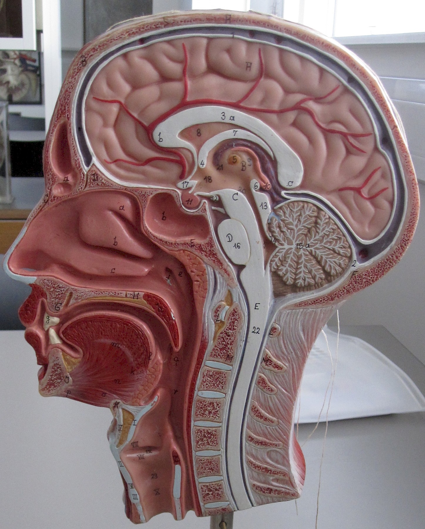 3D model for teaching anatomy. Source: Somso model from Adam,Rouilly. Photo by Elizabeth Hallam, 2014
Introduction
Drawing on anthropological research in medical school settings in Scotland, this article examines the contemporary anatomising of human bodies after death – that is, the anatomical dissection and study of bodies for the purposes of medical education – as a relational process. It considers three main aspects of this process: the social relations of the dead, namely, the social relations of the deceased who have donated their bodies to medical education; the relations between human bodies and the different media used in the teaching and learning of anatomy (for example, MRI scans, X-rays, computer-based anatomy software, and three-dimensional plastic models); and the anatomical relations that are examined and visualised within bodies as students develop their knowledge of anatomy. In contexts of medical education these relations are variously highlighted or, alternatively, de-emphasised and occluded, such that each body donor is constituted, over time, as a deceased person, an anonymous cadaver, ‘material’ for learning (Guide 2009-10, 16), and an instance of human anatomy. With reference to practices at the University of Aberdeen in northeast Scotland, which I began researching in 1999 (see Hallam 2016), I analyse the anatomising of bodies from the initial arrival of the recently deceased at the medical school, to their use in the dissecting room, and their subsequent memorialisation by family and friends when returned for burial or cremation. Throughout this process, and depending on the particular relations that are foregrounded, bodies after death are valued as persons, as material for the generation and communication of anatomical knowledge, and as gifts for the advancement of medical science. This constitution of meaning and value informs the perceived affective potential of human remains and the responses they evoke in the living.
Highlighting the notion of relational anatomy, I argue that analyses of anatomy that emphasise the effects of anatomical practices in terms of the fragmenting and objectifying of the human body need to be modified by studies that demonstrate how anatomical practices also produce relationships, modes of connection and integration on social, material, and conceptual levels. Historical studies of anatomy have tended to associate dissection predominantly with bodily fragmentation, but recent anthropological studies that similarly take up the issue of anatomical fragmentation are beginning to prompt an analytical reframing. Sharp (2000, 289), for example, links dissection with forms of scientific knowledge that ‘fragment the body with increasing regularity’ whilst also emphasising the importance of power relations and ‘body integrity’ (ibid., 288). [note 1] In her study of anatomy and surgery education, Prentice (2013, 24) asserts that the ‘fragmentation of bodies is the hallmark of biomedicine’, even as this education is effected through interactions between bodies (including dead bodies) and technologies that produce ‘bodily, social and relational ways of knowing’ (ibid., 15). Furthermore, discussing the sociality of human bodies, Lambert and McDonald (2009, 5) argue that, within the sciences, ‘acts of bodily fragmentation … are never confined solely to the biologically functional in their effects but inevitably entail the reformulation, reconstruction or reestablishment of social relations between persons and between human groups’. The intimate entwining of the social and the biological becomes evident in the research setting I describe below through a focus on relational anatomy, despite the work of differentiation that the practice of anatomy often performs with regard to these two domains.
In the next section I contextualise the notion of relational anatomy within anthropological debates and within wider institutional and technological environments. I then trace the social, material/visual, and anatomical relations as perceived and constituted in this field of practice. To conclude I note the transformations through which the dead move when dissected and memorialised; such transformations prompt questions with regard to dominant conceptions of the body currently generated by contemporary biomedical practices, and cut across assumed distinctions between dead and living bodies.
Relations: Debates and contexts
Analysing the relational aspects of anatomy as a contemporary field of knowledge practices that crucially involves deceased bodies – despite critiques of this involvement and the design of different pedagogical methods that aim to dispense with the dead – counters the emphasis on bodily parts and fragmentation in historical accounts of Western anatomy (see for example Cunningham 2010; Ferber and Wilde 2011; Hillman and Mazzio 1997). [note 2] In contrast to studies that highlight the importance of dismantling and partitioning bodies, I argue that in current processes of anatomising there is a dynamic interplay of separation and relation, of distance and proximity, which develops through medical school dissection and memorialising. Analysis of this interplay offers an anthropological perspective on the biomedical constitution of the human body not only as a clearly bounded or discrete individual physical unit, a pervasive effect commonly noted of Western biomedicine (see Lawton 2000; Lock and Nguyen 2010). This body is also a changing entity that is seen to be composed of intricate inner anatomical relations, it is understood through interrelated media used to display anatomy in medical education, and it is thoroughly enmeshed in wider social relations. These relations cut across distinctions between what is assumed to be clearly internal or external to the body as separated by the skin, the bodily organ that is typically thought to form the boundary of a person in Western conceptions (Farquhar and Lock 2007; Lock and Nguyen 2010). And these relations are, by turns, made powerfully manifest or de-emphasised when the dead are dissected by anatomists and medical students.
The anatomising of human bodies for anatomical education in medical schools, especially in the United Kingdom and the United States, has been subject to anthropological and sociological study for some time (see for example Becker et al. 1961; Good 1994; Hafferty 1991; Sinclair 1997). Taking dissecting rooms as the primary locus of attention, such studies explore the social and cultural significance of bodies after death. In these spaces, students’ intensive interaction with corpses has been interpreted as an initiation into the medical profession, an important aspect of their extended rite of passage from novice to expert. Through it students gain knowledge, experience, and status, undergoing a significant and emotionally charged education (Sinclair 1997). Indeed, as Good (1994, 72) points out, students ‘engage in reshaping their experiential world’, learning specialised vocabularies along with ways of seeing and acting that, it is argued, reconstitute persons – whether living patients or dead bodies – as ‘object[s] of medical attention’ (ibid., 73). While these studies interpret the practice of dissection as transformative for students, here I am concerned with the transformations undergone by the dead, which variously constitute bodies as deceased persons, ‘cadavers’, anatomical material, and as gifts to institutions that sustain and develop medical science (see Hallam 2005, 2007). Within what anatomists describe as the rapidly changing context of contemporary anatomy teaching in the UK, those who donate their bodies to medical schools are materialised as instances of human anatomy and then subsequently memorialised in ways that underline the public benefit derived from their personal body bequests. The social and material relations entailed and produced in these processes – which unfold not only in the dissecting room but in further medical school spaces, including museums and sites where memorials are located – guide responses to remains of the dead, including the attribution of value.
Bringing together debates in medical anthropology and anthropological studies of material culture (see Gell 1998; Gosden, Larson, and Petch 2007; Hallam 2016; Harvey and Knox 2014; Peers 2009) facilitates the interpretation of anatomical knowledge production as a relational process, one that necessarily involves the tracing and forming of connections as well as cutting, distancing, and separating. Three main interconnected domains of relations develop through contemporary anatomy teaching practices that involve close work with the dead: i) social relationships, especially those forged among body donors, anatomy teachers, technicians, and medical students (see Hallam 2005, 2007); ii) material and visual relationships, which emerge within a process of anatomical intermediality (see Hallam 2006, 2016), which is central to the dynamics of anatomy teaching as a hands-on participatory practice; and iii) relationships within the body that are examined and conceptualised by anatomists and medical students as integral to human anatomy (see Moore and Agur 2002). ‘Anatomical intermediality’ here refers to the relations that are seen, felt, described, conceptualised, and otherwise posited between material objects, images, and texts in the practice of anatomy, in this instance in the context of medical school teaching and learning (Hallam 2006, 2016). These three domains of relationships are instigated and formed through medical school practices that i) are guided by expectations and legal requirements regarding the appropriate treatment and disposal of the dead; ii) involve an array of changing teaching methods, aids, and technologies; and iii) engage in the production of knowledge of the human body that is empirically grounded and disciplined yet nevertheless imaginative. Setting a process of anatomical intermediality into motion through medical school teaching and learning enables the visualisation of human anatomy not as dead, static fragments but as integrated bodies that are alive, moving, and growing. Engagement with this intermediality, therefore, transforms the dead into embodied knowledge of the living for medical students, achieving a form of animation that, in some respects, is seen to make the dead live. [note 3]
Medical education is an embodied, sensory, and social process, involving specific modes of interaction with materials and objects. Recent research on anatomy teaching in medical school dissecting rooms in the United Kingdom and United States analyses the constitution of knowledge through the subtle sensory interplay of students’ learning bodies with bodies of the dead. This sensory engagement with the dead promotes heightened awareness of a range of different relationships. For example, Fountain (2014, 21) explores the embodied act of observation conducted by students at an American medical school as a process of ‘hypothesis confirmation’ in which they learn to recognise the ‘descriptive and relational evidence of the anatomical body’. ‘Relational evidence’ in this instance refers to the evidence that students seek when an anatomical structure is examined and identified in concert with nearby anatomical parts within the body, and when the relationship between a bodily structure and its function is studied. Thus, Fountain (2014, 98) argues that ‘recognition of the relational evidence of [anatomical] structures in question … entails analysis of the visual and haptic features on display in cadavers’. In this context, then, Fountain examines relations in terms of the embodied practices through which medical students come to recognise and to narrate physical relationships between the human body’s anatomical parts.
Referring to ‘relational ways of knowing’ – again in US medical schools – Prentice (2013, 15) describes how the sustained tactile interactions involved in dissection forge connections between student and cadaver; the student’s understanding of the cadaver’s anatomy is shaped and enhanced through the ‘mutual articulation’ of the learner’s body and the body of the deceased undergoing dissection (ibid., 71). With similar attention to relationships, McDonald (2014, 129, 131, 135) – researching both anatomy teaching in UK medical school dissecting rooms as well as surgery and organ transplants – explores how students learn to recognise ‘relational parts’ of the body by studying in a ‘learning environment’ where multiple ‘anatomical bodies’ in different media (for example, atlases, manuals, skeletons, and cadavers) are assembled and undergo a ‘matching up’ or ‘reconciling’ (see also Mol 2002). According to McDonald, by learning skills in such an environment, students acquire a ‘medical body’ with abilities to see, handle, and understand anatomy, and they acquire this skilled body ‘relationally’, along with the other students, cadavers, artefacts, and instruments involved in this process within the dissecting room (McDonald 2014, 134; see also Olejaz, this issue).
In the sections to follow I develop the analysis of how anatomical knowledge is generated and communicated by further examining the social, bodily, conceptual, and material relationships that guide, and are produced through, anatomical practices. This relational anatomy, as I will argue, features a dynamic intermediality – which is significant, yet underexplored, in transformations of the dead into knowledge of the living body (Hallam 2006, 2016). Anatomy teaching and learning proceeds through situated material and visual practices that shape the bodies of those involved, including medical students and the deceased; and these processes are caught up in (and productive of) relations that become particularly salient for the participants engaged in this knowledge-generating work.
How relations are constituted and understood in the making of expert knowledge is explored in Rival’s (2014) discussion of ecology. She acknowledges extensive anthropological debate on relationality – including work by Marilyn Strathern, Tim Ingold, and Philippe Descola that offers different formulations of the concept of ‘relation’ – whilst also underlining the need to examine the specific contexts and knowledge-making practices in which this concept is variously mobilised and attributed meaning. In a similar vein, I am concerned with particular kinds of relations that are constituted, reinforced, or occluded in one field of biomedical science – that is, in anatomical practice – especially as they pertain to the dead. Human remains, preserved for the purposes of anatomy, are relational entities whose forms and material properties emerge through embodied interactions that take place with them, interactions that are inescapably situated within particular fields of social and power relations (Hallam 2010, 2016). This relationality by no means excludes modes of separation, especially as the cutting and dismantling of bodies, for instance, has been so crucial to anatomy in practice. Indeed, current anthropological work concerned with the ‘limits of relational thinking’ points to the interpenetration or co-implication of separation and relation, so that Candea and coauthors (2015, 16) draw attention – via discussion of Strathern’s and Latour’s work in particular – to ‘the ways in which disconnection, cuts and distinctions, accompany, undergird, permit or arise from relations and connections’ (see also Myhre 2016). Ethnographic study enables analysis of diverse processes through which relations and separations, engagement and distancing, variously play out in practice. The cultivation of distance, dispassion, and objectivity in anatomy has a complex history (see for example Daston and Galison 2007; Payne 2007) as do the relations in which anatomical work is enmeshed (Hallam 2016), and in current medical school dissecting rooms detachment is no less a nuanced, negotiated process in the learning of anatomy (see McDonald 2015).
Contemporary uses and treatment of deceased body donors in medical schools, it is important to note, are not undertaken in isolation but rather within wider changing institutional and technological environments, which I briefly outline here. Practitioners involved in teaching anatomy have noted, with reference to the United Kingdom in the last twenty years, a decline in teaching staff, rising numbers of students, and a reduction in the number of hours dedicated to anatomy in the medical curriculum – a cut, some argue, that might lead to unsafe clinical practice (see Turney 2007). Anatomists and medical educators draw attention to a decline in human dissection since the 1980s, and also to variation in teaching methods across different medical schools (Older 2004; Turney 2007; Davis et al. 2014). In some schools students undertake dissection, in others students learn with prosections (preserved body parts already dissected by staff), and at one school that opened in 2002 there is no use of deceased bodies (McLachlan et al. 2004). Another school reports that students are taught with plastinated prosections, purchased from Von Hagens Plastination, in Guben, Germany (Fruhstorfer et al. 2011). These ‘plastinates’ are ‘manufactured’ from body donors, which restricts sales of prepared body parts to ‘institutions or individuals who use specimens exclusively for research and educational purposes or for medical, diagnostic and therapeutic education’. [note 4]
Medical students also learn from living models (themselves and professional life models), and with visual images of live bodies produced through medical imaging techniques, especially radiography, computed tomography (CT), and magnetic resonance imaging (MRI), which are increasingly used in clinical settings. Furthermore, there has been a rise in the use of plastic models and of anatomy software (Older 2004). Computer-based anatomical images include those derived from bodies of the dead, such as the digital images from the U.S. National Library of Medicine’s Visible Human Project, initiated in the 1980s to create a ‘digital image library of volumetric data representing complete, normal adult male and female anatomy’ (NLM 1996, see also Dijck 2005; Donnelly et al. 2009; Waldby 2000), and the ‘virtual cadavers’ composed from multiple CT scans and combined with artists’ digitally rendered images, as in the commercially available life-size Anatomage Table, which allows touch-interactive virtual dissection. The Anatomage Table is designed and marketed by Anatomage, a company specialising in 3D medical technology in California, in collaboration with Stanford University’s Division of Clinical Anatomy. St Mary’s Hospital in London purchased the first to be used by a medical school in Europe in 2012. [note 5] The pedagogical resources under development for anatomy teaching in the UK range from the high tech, as in three-dimensional computer-generated images and models produced with 3D printing technology, to the low tech, as in anatomical model-making from electrical wire and other easily accessible materials (Hallam 2013a; Lim et al. 2015; Thomas 2010; Trelease 2016). Anatomy teachers have used PVC-coated wire, for instance, to interactively model nerves in situations where medical students find certain three-dimensional pathways of nerves through particular parts of the body difficult to visualise.
The General Medical Council (GMC), which sets requirements for UK medical education and training, and issues the licenses that allow doctors to practise, does not specify that students must dissect or study with deceased bodies. It does, however, require them to gain ‘practical experience’, learning from patients in clinical settings and through the provision of ‘access to technology enhanced and simulation based learning opportunities’ (GMC 2015, 34). Each medical school is expected, then, to design its own curriculum in accordance with the GMC’s standards, yet within these guidelines there is latitude, as medical school staff note that ‘anatomy teaching is approached differently according to departmental culture’ (Sugand, Abrahams, and Khurana 2010, 84). While the GMC’s recommendations place emphasis on learning with living bodies (of patients) and with technology, the Anatomical Society – the UK’s main learned society for promoting anatomical science, whose members are mainly those involved in research and teaching in higher education – emphasises that ‘the study of the dissected human body remains the “gold standard”’ in methods for gaining anatomical knowledge (Anatomical Society, n.d.). This society also outlines a ‘core of anatomical knowledge that will equip students in the UK and Ireland for safe and effective clinical practice’ (McHanwell et al. 2007, 18), which should be attained ‘whatever teaching approaches are adopted within a curriculum’ (ibid., 4). How best to teach and learn anatomy continues to be debated by anatomists and medical educators in professional organisations and specialist journals. [note 6] But a common feature of all medical schools appears to be their use of combinations of materials (see Johnson, Charchanti, and Troupis 2012; Mitchell and Stephens 2004) – as no single medium for teaching anatomy is deemed sufficient in itself – and it is in relation to these specific combinations, selected and brought into play in each local teaching situation, that bodies after death are interpreted and attributed pedagogical value.
The meanings that deceased bodies accrue in anatomy teaching are also shaped by a wider social context where there are strong public expectations with regard to the appropriate treatment and disposal of human remains by the medical profession. Sensitivity to these issues was heightened during the hospital inquiries that revealed large-scale organ retention from children’s postmortems in 2000–2001 at Bristol Royal Infirmary and the Royal Liverpool Children’s Hospital (Alder Hay). The new legislation that followed, the Human Tissue Act of 2004 (England, Wales, and Northern Ireland) and the Human Tissue (Scotland) Act 2006 sought to ensure the informed consent of donors and tighter licensed control of the procurement, use, and disposal of human tissue (see Jones and Whitaker 2009). Although it has been observed that these scandals, widely commented on in the media, seem not to have discouraged people from donating their bodies to medical schools (see Wilton 2007), numbers of body bequests continue to fluctuate; there was media reporting of declining donations in 2006 then subsequent rises in 2007 and 2010 (see de Bere and Petersen 2006). [note 7]
Medical schools that teach with deceased bodies are, therefore, keen to publicly stress the importance and value of donations, while also highlighting the kind of care that bodies receive. Calls for bequests on medical school websites emphasise that donations will be appropriately cared for; King’s College London, for example, whose bequests are dealt with by the London Anatomy Office, emphasises that bodies are treated with respect and that medical students are encouraged to treat bodies ‘suitably reverentially’ [note 8] (see Douglas-Jones, this issue). This discourse and practice of appreciation and decorum is ritually reinforced in the annual memorial services for donors and dedicated memorials that have increased since the 1980s (Druce and Johnson 1994; Tinker 1998). Such services are currently held, for instance, by medical schools in Aberdeen, Bristol, Cambridge, Glasgow, Keele, and London. The University of St Andrews’ new medical school building (opened in 2010) has a book of remembrance for donors exhibited in a display case that is publicly viewable, in 2012 Keele University established a memorial stone for donors in a local cemetery, [note 9] and in the same year Cardiff’s School of Biosciences installed a memorial for donors to be viewed by medical students (rather than the public) in the anatomy laboratory. Designed by artist Tom Philips, the memorial – inscribed ‘Alive we thought beyond our lives to give our bodies as a book for you to read’ – gives a voice to donors after death and reminds students, according to anatomy professor Bernard Moxham, that as their first patient each body donor is a person who has shown an enormous ‘generosity of human spirit’ that makes the work of anatomy possible. [note 10] Expansion in medical school memorial events, with their own local practices and messages about deceased body donation, is also apparent in the Netherlands (Bolt 2012); the United States (see Fountain 2014; Jones, Lachman and Pawlina 2014; Prentice 2013); Melbourne, Australia; Cape Town, South Africa; Hualien, Taiwan (see Douglas-Jones, this issue); and Phitsanulok, Thailand (Labuschagne and Mathey 2000; Lin et al. 2009; Winkelmann and Güldner 2004). [note 11] There is, furthermore, increasing (potentially) worldwide visibility of donor memorials on the internet, as medical schools in the United States, for example, use websites such as YouTube to publicise them. [note 12]
These two broad developments – changes in medical school teaching with an accompanying expansion in the range of educational anatomical materials, and the growth in memorials for body donors – construct deceased bodies in particular ways. Significantly, they shape perceptions of the dead as relational entities with both potent material dimensions and specific attributes and affects as people. The rest of this article analyses this relational formation and transformation of deceased bodies, with attention to the social and the anatomical relations involved. The discussion is based on research beginning in 1999 (and still ongoing) at the Anatomy Department of the University of Aberdeen, where I have conducted ethnographic, archival, and museum-based research in parallel with research at additional medical schools in Scotland and England. The department was based at Marischal College until 2009, when it moved to new purpose-built premises in the Suttie Centre, at Foresterhill Campus, forming the Anatomy Facility in the School of Medicine and Dentistry (now the school of Medicine, Medical Sciences, and Nutrition) located with Aberdeen’s hospitals. The following analysis relates to anatomical practices up to the time of this move, although many of the teaching and memorial practices I discuss are still ongoing and are the subject of my research visits since 2010 (see for example Hallam 2013a, 2013b). As a member of the university staff (in the Anthropology Department until 2010), I regularly visited the Anatomy Department to talk with and observe anatomy teachers, technicians, and students at work in the dissecting room, the anatomy museum, staff offices, storage areas for teaching materials, and other spaces. [note 13] I interviewed retired members of staff, worked with a professional photographer to document aspects of the museum and the collection of teaching materials, especially 3D models, and undertook a detailed study of historical documents kept in the department’s archive room. The analysis below arises out of this in-depth research, and I provide more detailed descriptions in further recent explorations of anatomical practices (see especially Hallam 2016).
Social relations of the dead 1: Distance and anonymity
Transported to the Anatomy Department in Aberdeen no more than a few days after they had died, the bodies of donors were delivered by undertakers to the mortuary located in an area with highly restricted access. This was a sub-basement two floors below the dissecting room. In order to be accepted, donors would have completed the essential forms, consenting in writing to their body being ‘made available for anatomical teaching, training and research’ for up to three years (or optionally longer). [note 14] Donations were managed by the anatomy bequest administrator; she received enquiries from potential donors, most of whom lived in the north of Scotland, kept an archive of donor bequest forms (which began in 1965), organised the delivery of twenty to thirty deceased donors’ bodies per year to the department, and made arrangements for donors’ subsequent funeral and memorial services. The administrator was thus a central figure in the ongoing social relations of donation among donors, next-of-kin, executors, medical practitioners, and funeral directors, among others. Her work at the interface of anatomy teaching and body donation by members of the public was key. She participated in matters at once legal and personal, bureaucratic and emotional, mundane and ritualised.
In this setting, donors registered their intention to donate between four and thirty-four years (on average sixteen years) before they died, and at death donors ranged in age from their late fifties to 100 years old (the minimum age is seventeen years). [note 15] Family members often made intended bequests together, for example when married couples both wanted the same method of disposal after death. Furthermore, in such cases the family intention for such donations to be carried out could lead to disappointment on the part of surviving relatives if only one of the intending donors was accepted in practice. Richardson (1995) suggests that having known someone who was accepted as a donor or who aimed to donate was a significant factor in many decisions to make a bequest to medical schools in London during the 1990s. If bequests are generated through social connections, the acceptance of bequests depends on the donor’s physical condition at death; medical schools specify, for instance, that donors should not have donated an organ other than corneal transplant, or had a postmortem examination/autopsy, recent major surgery, a history of certain infectious diseases, obesity, advanced cancer, or some kinds of dementia. [note 16] The acceptance of deceased bodies offered by donors while alive, then, depends on a number of intersecting factors at the point of death, many of which cannot be anticipated beforehand, and some of which rest on material requirements for managing bodies of the dead in concert with socially and culturally defined priorities regarding the selection of bodies for anatomical education.
Once in the mortuary, two technicians prepared a donor’s body for use in teaching. From their perspective, the deceased donors, arriving so recently after dying, were still strongly enmeshed in the relationships they sustained during life. The junior technician, who began in 2004 as a young trainee, explained to me that when a donor was first laid out on the embalming table – still in their clothing, usually nightwear or hospital wear – she was very aware of them as a person, unknown to her but whom she imagined had relatives ‘now left behind’. She found herself treating donors almost as though they were still alive, yet the mortuary procedures she performed with care and respect tended also to distance (but not absolutely extract) donors from their former social lives. A donor’s name was recorded in a logbook and they were assigned a unique number; this replaced the person’s name to maintain a donor’s privacy in the Anatomy Department. But to anonymise in this way also removed a marker of personhood and social relatedness (see vom Bruck and Bodenhorn, 2006). Next, clothing was removed from the donor’s body and any jewellery they were wearing was given back to their next-of-kin. After washing, disinfecting, and shaving, the embalming of the donor began to ‘prevent decomposition and the growth of microorganisms’; bodily fluids, including blood, were drained and preserving fluid was pumped through the vessels (Guide 2009-10, 14). Tags bearing the donor’s number were then attached to ensure clear identification of all body parts that might become separated during dissection.
Through these procedures the donor as person also became what was referred to in this context – by staff and students – as an ‘embalmed subject’, a ‘body’, a ‘cadaver’. This was a material as well as a social transformation (see Hallam 2007). Embalming for instance, hardens tissues and changes body colour to muted shades of beige and grey, giving the cadaver a pale, almost waxy appearance. The junior technician observed that with these changes the physicality of the dead body became more pronounced, as the once-living appearance of the deceased person seemed to recede. McDonald (2014, 138) suggests, with reference to her anthropological research on dissection, that when ‘inherently’ social bodies are anonymised during their admission into medical schools, this ‘anonymity severs relations’. I would argue, however, that in this context, anonymisation did not sever previous social relations, especially as material traces of a person and their life – and therefore of their social connectedness – were perceived to remain on and within their embalmed body. So, significantly, the personhood of donors along with their social relatedness was not entirely extinguished; rather, it remained latent, so that bodily scars and marks, for example, were seen by staff and students as indicators of the deceased’s former life and relationships. Looking at some donors’ hands, for instance, the teaching fellow interpreted nail polish as a reassuring sign that female donors had been cared for at the end of life. In the case of this cosmetic, associated with personal care and the aesthetics of appearance while alive, nail polish on the hands of the dead had the capacity to invoke sensations of connection with, and compassion for, body donors as people with social lives (see also Levin 2000, 15; Montross 2008, 24; Prentice 2013, 45). Furthermore, donors as persons were also apparent to medical students as they worked in the dissecting room, which I discuss next.
Anatomical relations 1: Materials and display
Distanced from their previous social relations, in the dissecting room donors as cadavers entered into a new set of relations through practices of teaching and learning anatomy. Prior to 2003 students would dissect a body in groups of about eight, at approximately thirty steel dissecting tables, guided by teaching staff. Dissecting in ‘anatomical regions’ – the head/neck, thorax, abdomen, upper and lower limbs, back, pelvis/perineum – students removed skin and fat, cutting progressively deeper to reveal the arrangement of muscles, organs, blood vessels, nerves, and bones. The work of opening, cutting, and separating was done in order to see and feel anatomical parts and their relations; material cutting in this process made anatomical relations knowable. By enacting these practices students rendered the cadaver’s anatomy visible and intelligible. But in doing so they encountered difficulties, especially with regard to their perceptions of a cadaver as a person. When students found scars of childbirth or surgery in cadavers, for example, these were seen and felt as physical evocations of those bodies’ personal histories, as physical signs of lives lived. When these sometimes emotionally moving impressions made dissection especially troubling, students learned to distance the person from the cadaver they were studying; they learned to manage their perceptual oscillation between sensing the cadaver as person and as embalmed body. Further studies of medical school dissection comment on a similar process; Prentice notes that students in the United States develop a strategic ability to ‘objectify the body’ on the one hand and, alternatively, to ‘call forth the person’ on the other. She describes this movement as ‘tactical objectification’ (Prentice 2013, 35). The relation between person and body is, therefore, not disconnected; rather, the two are moved in and out of focus as students place themselves in relations of either distance or proximity vis-à-vis the deceased body. Fountain (2014), discussing US medical schools, similarly identifies an interplay or balancing of detachment, concern, and empathy (see also McDonald 2015; Olejaz 2015).
At the start of dissection classes, anatomy teachers in Aberdeen impressed upon students the generosity of donors and the expectation that they would ‘pay’ their ‘respects’ by attending the memorial service at the end of the year. A code of conduct in the dissecting room, including rules for the ‘care of cadavers’ was strictly enforced and respected. Within this framework, donors were treated as valued anatomical ‘material’. Through interaction with this material, students were advised that they must develop the skills of ‘being able to visualise in the mind’s eye and feel with an examining hand the body structures as they lie beneath the skin’. Intensive training of students’ eyes and hands was meant to enhance their capacity to visualise in three dimensions ‘how parts of the body are put together and how these components work’ (Guide 2009-10, 5–10).
To facilitate this learning, cadavers were dissected and examined among an array of further anatomical materials, in comparison with which they were defined as the most pedagogically effective. This evaluation of cadavers’ powerful educational efficacy remained the case, even when the department replaced dissection with prosections in 2003, so that with this change students handled and examined preserved body parts already dissected by staff (prosections) instead of conducting dissection themselves. Cadaveric material was described as more ‘real’ than any other material; ‘this is as real as it gets’, said one student. With cadavers, students could see variations in different bodies and they could feel the shape and texture of anatomical parts in situ. But while considered powerfully ‘real’, cadavers were clearly recognised as differing from living bodies. Therefore, in the dissection room, students had to imaginatively extrapolate from the dead to the living. They were told: ‘anatomy is the people around you, alive and moving, from the vigorous flexing of muscles to the gentle peristaltic movements of the intestines… . Remember our anatomy is alive and moving not dead and static’ (Learning Guide 2006-7, 4–9).
To enable students to visualise living anatomy from the dead – or to imaginatively transform inert bodies into animated ones – a wide range of anatomical materials was displayed, in the dissecting room, the museum, and other teaching spaces. [note 17] Plastic models as well as X-rays, CT, and MRI scans from anonymous live clinical cases were mobilised (see Fountain 2014; McDonald 2014; Prentice 2013). Students also studied with commercial anatomy software at computer stations, such as Acland’s DVD Atlas of Human Anatomy, which allows students to observe images of dissected body parts that are unembalmed and so claim closer resemblance to the living body. Significantly, students’ own bodies were also utilised in surface anatomy classes; some voluntarily acted as living models and, in small groups, students would palpate each others’ muscles and draw on skin with cosmetic crayons to indicate the position and shape of organs. Students defined themselves as anatomical material when they were encouraged to compare parts of dissected cadavers with corresponding parts of their own bodies’ surface anatomy; this linkage of dead and living bodies helped students to visualise the anatomy of the former in terms of the latter, in order to animate or bring it to life, an issue to which I return in the conclusion.
Students learnt to ‘correlate’ (link and compare) these materials, to see and feel relations between them as a means to clarify and enhance their anatomical knowledge of living anatomy (Learning Guide 2006-07, 5). For example the ‘same’ aspects of the anatomy of the head were examined in the cadaver, through plastic models and MRI scans. This was an intermedial process in which any number of body parts, live and deceased and distributed across different media, were brought into meaningful relation in the process of learning (Hallam 2006, 2016). I saw manifold instances of this anatomical intermediality in the Anatomy Department due to its centrality in the teaching and learning process. For example, when studying the head and neck in the dissecting room, one student visited the adjacent Anatomy Museum where, on a table, she gathered several 3D plastic models of the head and brain that she studied in relation to diagrams in a textbook and her own notes and sketches (Hallam 2016). Crucial in this learning practice was the development of the capacity to navigate fluently between 2D images and 3D renderings of anatomical parts (such as X-rays, prosections, and aspects of surface anatomy), to interpret in depth the complex spatial relations of those parts. The next section examines these relations.
Anatomical relations 2: Inside bodies
By tracing relations between these materials, then, students explored anatomical relations deep within bodies. One textbook guiding students’ learning, therefore, highlighted the ‘relationships of various systemic structures (e.g. muscles, nerves, and arteries) within the region, part or division of the body’ (Moore and Agur 2002, 2). While anatomical parts were identified and named, anatomy was not understood in terms of isolated parts because these had to be situated within the body, understood as related structures envisaged within living bodies. So, numerous ‘key relationships’ in each region of the body – for example, in the abdomen, those of the oesophagus, stomach, and small and large intestines – were listed as integral to the ‘core knowledge’ outlined by the Anatomical Society (McHanwell et al. 2007, 4, 8; see Thomas et al. 2010). Studying these relations was deemed to produce ‘factual knowledge’ of already-existing organic anatomical structures that were present and largely independent of the learner (McHanwell et al. 2007, 4). Nevertheless, the conception of knowledge operative here, which was shared by both teachers and students, was one that required a close relationship between cadaver and student. To learn anatomy as a system of relations, students had to form close interconnections with cadavers through practical study.
Learning anatomy is a visual education requiring disciplined bodily action; it is an embodied process during which a student takes visual and tactile impressions from anatomical materials in order to thoroughly internalise them. Students were taught that this learning ‘build[s] up a complete 3D image in your mind’, which mentally assembles and integrates ‘pieces’ of the body into a functioning ‘single unit’ (Learning Guide 2006-07, 11). This 3D image is envisaged as dynamic and cumulative: it develops over time as the student interacts with anatomical materials during their training and it will be further augmented through their future medical practice. Teachers, with years of experience, are seen to possess ‘expert 3D conceptualisations’ compared with those of novices, which are still to properly form (Patten 2007, 14). These teachers help students actively participate in developing their own understanding of these ‘complex three-dimensional … relationships within the human body’ (Patten 2007, 10). Publications in international journals dedicated to medical education further reinforce and circulate this approach to anatomy teaching and learning: ‘Mastering anatomy requires understanding of three-dimensional relationships. Students expend considerable effort not only to learn orientation, location, and structural dimension, but also to retain this information. Failure to master and retain an accurate mental model of these concepts can negatively affect success in clinical and academic practice’ (Bareither et al. 2013, 170).
Such 3D mental images are neither entirely derived from anatomical materials, nor entirely imagined, but continually emerge somewhere in between. Thus this process of knowledge formation undermines easy distinctions between the material and the mental, person and thing, the living and the dead, the organic and the artificial. Students’ mental images of the anatomical body are composed through interactions with a range of anatomical material so that impressions of dissected cadavers are integrated with those of plastic models, digital and clinical images, diagrams, and their own bodies. A student’s embodied impressions of donors’ bodies are thus incorporated as anatomical knowledge (see Connerton 1989). The cadaver is transformed into embodied knowledge, forming part of the student’s sense of their own internal self. The intimacy and significance of this relation between students and cadavers, formed through learning, was felt and expressed by students at the Anatomy Department’s annual memorial service for body donors, which is still held each year. With regard to this relationship, a student’s views at Keele’s medical school are telling: ‘While you spend a year working with the body, you are looking into this person’s life, how they lived it, and things they went through. By the end of the year it has become a very personal relationship’. [note 18] Aspects of that relationship were brought to the fore at Aberdeen’s annual memorial service for body donors to the medical school.
Social relations of the dead 2: Remembering and memorialising
At the end of the allocated period of time for donors in the Anatomy Department (a maximum of three years), their bodies were prepared for removal by the technicians, supervised by the senior anatomy lecturer. Each dissected cadaver was reassembled in a coffin in the dissecting room. This re-membering of each body brought together parts separated during dissection and prosection, yet this would not form the donor’s ‘whole’ body: for example, blood had already been removed and fat would have melted and dispersed during dissection, but at least two-thirds of the body would still be sent for cremation or burial. This radically altered body was, then, removed from the social relations of the Anatomy Department, and extracted from a nexus of relations among the array of anatomical materials used in teaching, to receive ritualised disposal and memorialising. Unlike the nameless dissected bodies in the anatomy software that students view, which remained suspended in their digital afterlives, the numbered bodies of donors were reunited with their names and their personhood brought to the fore through memorials that re-embedded donors in their former social relations among family and friends (see Hallam 2007).
The anatomy bequests administrator was responsible for co-ordinating the donor’s funeral arrangements and memorial service. She instructed funeral directors to transport donors to Aberdeen Crematorium or, in one or two cases, to nearby Trinity Cemetery for burial, according the donor’s wishes (unless next-of-kin were arranging a private funeral). A larger memorial service, led by the chaplain to Aberdeen University, was held every May at King’s College Chapel to thank and remember those people who had donated their bodies during the previous year. [note 19] The service, which is still held to date, was initiated in the 1950s, the first of its kind in Scotland, and possibly Britain (and held in the dissecting room until 1972). Over the last decade or so, the memorial service has brought together, each year, about three hundred attendees, including donors’ relatives and friends, anatomy teachers, and medical students. Providing a focus for grief, the emotional high point of the occasion is the reading of the donors’ names, the name being a key point of attention at other memorial events for body donors held by medical schools in Britain (see Tinker 1998). For attendees who knew a donor when alive, the name can evoke moving memories of the deceased and their past life. For students, hearing donors’ names for the first time reinforces their sense of dissected and prosected bodies as persons connected with living people who still keenly remember and care for them. Students witness relatives’ expressions of personal loss, potentially increasing students’ sensitivity and empathy in situations surrounding death. In these ways the ritualised social reintegration of the deceased establishes connections between medical students and the donors whose bodies they have learnt from and whose relatives and friends they join at the memorial service.
On display at every service is the memorial book, inscribed with each donor’s name. Begun in 1966, this has been kept, protected, in a locked glass case in the Anatomy Department with an accompanying inscribed plaque: ‘This book records in gratitude the names of those who bequeathed their bodies for the advancement of medical science’. The inscription of all donors’ names in the book registers respect and gratitude from anatomists and students, while the plaque associates body donation with a wider culturally valued process: the ‘advancement of medical science’. The linkage of donors’ body bequests with medical advancement is made on a further memorial in Trinity Cemetery, built in 1974 (again probably the first in Britain) on a large plot where dissected bodies have been buried since 1921 in unmarked graves. Facing the cemetery, the memorial’s dedication reads: ‘In memory of those who gave their bodies for the increase of knowledge and the advance of medicine’.Deliberately minimal, in concrete and stone, this collective memorial is devoid of names; rather it marks a collective, united in giving their bodies for the benefit of others. A second inscription announces: ‘Their name liveth for ever. The people will tell of their wisdom and the congregation will show forth their praise. Ecclesiasticus 44’. A further memorial stone for body donors was installed in 2007 in the grounds of Aberdeen Crematorium, carrying the same inscription as that at Trinity Cemetery. The predominant message conveyed by these related memorials is that death, or more precisely the dead body, can be transformed into life-giving medical knowledge to the benefit of all.
These publicly visible interrelated memorials are simultaneously expressions of appreciation for donors’ gifts, invitations to further donors, and statements of medical progress. Memorials operate as mediators, establishing and maintaining relations between anatomy teaching staff, donors and their families, as well as potential future donors (see also Fountain 2014). Such memorials are made to participate in a powerful public discourse that suggests body donors, through their common ‘sacrifice’, enter a community of the dead that contributes to the maintenance of health and therefore the improvement and extension of human life. Memorial inscriptions for deceased body donors also resonate with war memorial inscriptions (which use the similar quotations) for those who ‘gave their lives’, highlighting sacrifices made for others (see also Lock 2002; Sharp 2006). Such invocations of sacrifice reinforce one of the paramount values in biomedicine, namely the maintenance of human life and the reduction of suffering, which, as Good (1994, 86) has argued, medical work and discourses highlight as central aims made possible by the immense and seemingly ever-advancing ‘technical efficacy of medicine’. Bodies after death are thus constituted as crucial resources that sustain and enhance that efficacy or capacity to save lives and improve life.
Transformations of the dead
As each deceased body donor in Aberdeen underwent the process of anatomising, their body after death was sequentially transformed into a cadaver, anatomical material, students’ 3D mental images, a person to be remembered, and an agent in the medical saving of lives. These transformations were brought about in sites of anatomical practice, including memorials, that were both sustained by, and generative of, particular kinds of social, material, and conceptual relations. Value was assigned to donors’ bodies within these relations, which also guided affective responses to those bodies. Deceased bodies were viewed and manually explored as a system of anatomical relations in an institutional context that also promoted donors’ memorialisation as persons within a community of students and among the family and friends of the deceased.
To analyse anatomical practices in this way highlights the perception and constitution of deceased bodies as relational entities. This complicates views of anatomy as a predominantly fragmenting process. While human bodies are divided, cut, and separated – into regions and parts, for example – these divisions are performed and explored by students as routes towards the learning of anatomical relations. Division is not an end in itself but rather a phase in a process that generates knowledge of the body as integrated anatomical relations (rather than disconnected parts). In this way, dead bodies are transformed into knowledge of the living body; the dead are visualised as anatomically alive. Such an animation is crucial in converting remains of the dead into valued medical knowledge. A key aspect of this animation is the process of intermediality, in which students trace relationships between anatomical parts rendered in different media, from dissected bodies to printed diagrams, 3D plastic models, MRI scans, and students’ own bodies. This contextually situated tracing of relationships transforms dead bodies into students’ embodied anatomical knowledge of the living. The anatomical relations dissected and examined through disciplined sensory interactions with the dead are thus incorporated into the bodies of living students to provide the epistemological foundation of their future professional medical work.
If practices of anatomy have participated in the constitution of the human body/person as a ‘skin-bounded … biomechanical entity’ (Farquhar and Lock 2007, 2) – understood as a dominant Western conception reinforced by anatomical knowledge developing from the enlightenment onwards (Lock and Nguyen 2010, 40) – departures from (or additions to) this conception are also being produced in contemporary anatomical practices. As McDonald (2014, 136) has observed in medical school teaching, students’ participation in dissection prompts each to learn where boundaries are and to thereby acquire an ‘autonomous, bounded body-self’. In many ways the bodies of deceased donors are also treated and respected as distinct bodies – indeed, as noted above, each one is visualised as an integrated system of anatomical relations functioning within the body described as a discrete, ‘single unit’. But these bounded bodies are also constituted as relational: their anatomy is interconnected (socially, materially, and conceptually enmeshed) in important ways, especially as the living learn from the dead by incorporating aspects of them via embodied practices in medical school settings. This relational anatomy is a mode of animation that is dynamic and changing: it promotes visualisation of the dead as alive, it imbues the living with incorporated aspects of the dead (which take the form of anatomical knowledge), and it is central to practices in medical education that drive such transformations in the name of saving and improving lives.
Acknowledgements
I thank staff and students in the Anatomy Department at the University of Aberdeen for their invaluable help with my research. Many thanks also to Rachel Douglas-Jones and Bob Simpson, colleagues and friends who have generously encouraged me to develop this article especially through their inspiring workshop and conference meetings. I am grateful to two anonymous reviewers for their insightful comments.
About the author
Elizabeth Hallam, PhD, is Research Associate in the School of Anthropology and Museum Ethnography, University of Oxford, and Honorary Senior Research Fellow at the Department of Anthropology, University of Aberdeen. Her research and publications focus on the anthropology of the body, death and dying, material and visual cultures, histories of collecting and museums, the anthropology of anatomy, three-dimensional modelling, and mixed-media sculpture. Her recent books include Making and Growing: Anthropological Studies of Organisms and Artefacts (co-edited with Tim Ingold, Ashgate, 2014), Designing Bodies: Models of Human Anatomy from Wax to Plastics (edited, Royal College of Surgeons of England, 2015), and her monograph Anatomy Museum: Death and the Body Displayed (Reaktion, 2016).
References
Anatomical Society. n.d. ‘Body Bequeathal’. https://www.anatsoc.org.uk/about-us/bequests/information-about-body-donation.
Bareither, Mary Lou, Vered Arbel, Meghan Growe, Emily Muszczynski, Adam Rudd, and Jane R. Marone. 2013. ‘Clay Modeling versus Written Modules as Effective Interventions in Understanding Human Anatomy’. Anatomical Sciences Education 6, no. 3: 170–76. https://doi.org/10.1002/ase.1321.
Becker, Howard Saul, Blanche Geer, Everett C. Hughes, and Anselm Strauss. 1961. Boys in White: Student Culture in Medical School. Chicago: University of Chicago Press.
Bolt, Sophie. 2012. ‘When I Die I Will Go to the University: A Study of Body Donation in the Netherlands’. PhD diss., Radboud University Nijmegen and the Netherlands Organization for Scientific Research.
Candea, Matei, Joanna Cook, Catherine Trundle, and Thomas Yarrow. 2015. ‘Introduction: Reconsidering Detachment’. In Detachment: Essays on the Limits of Relational Thinking, edited by Matei Candea, Joanna Cook, Catherine Trundle, and Thomas Yarrow, 1–31. Manchester: Manchester University Press.
Carsten, Janet. 2011. ‘Substance and Relationality: Blood in Contexts’. Annual Review of Anthropology 40: 19–35. https://doi.org/10.1146/annurev.anthro.012809.105000.
Connerton, Paul. 1989. How Societies Remember. Cambridge: Cambridge University Press.
Cunningham, Andrew. 2010. The Anatomist Anatomis’d: An Experimental Discipline in Enlightenment Europe. Farnham: Ashgate.
Daston, Lorraine, and Peter Galison. 2007. Objectivity. Brooklyn, NY: Zone Books.
Davis, Christopher R., Anthony S. Bates, Harold Ellis, and Alice M. Roberts. 2014. ‘Human Anatomy: Let the Students Tell Us How to Teach’. Anatomical Sciences Education 7, no. 4: 262–72. https://doi.org/10.1002/ase.1424.
de Bere, Sam Regan, and Alan Petersen. 2006. ‘Out of the Dissecting Room: News Media Portrayal of Human Anatomy Teaching and Research’. Social Science and Medicine 63, no. 1: 76–88. https://doi.org/10.1016/j.socscimed.2005.12.011.
Donnelly, Leo, Debra Pattern, Pamela White, and Gabrielle Finn. 2009. ‘Virtual Human Dissector as a Learning Tool for Studying Cross-Sectional Anatomy’. Medical Teacher 31, no. 6: 553–55. https://doi.org/10.1080/01421590802512953.
Druce, Maralyn, and Martin H. Johnson. 1994. ‘Human Dissection and Attitudes of Preclinical Students to Death and Bereavement’. Clinical Anatomy 7, no. 1: 42–49. https://doi.org/ 10.1002/ca.980070108.
Farquhar, Judith, and Margaret Lock. 2007. ‘Introduction’. In Beyond the Body Proper: Reading the Anthropology of Material Life, edited by Margaret Lock and Judith Farquhar, 1–18. Durham, NC: Duke University Press.
Ferber, Sarah, and Sally Wilde, eds. 2011. The Body Divided: Human Beings and Human Material in Modern Medical History. Farnham: Ashgate.
Fountain, T. Kenny. 2014. Rhetoric in the Flesh: Trained Vision, Technical Expertise, and the Gross Anatomy Lab. London: Routledge.
Fruhstorfer, Birgit H., Julie Palmer, Stephen Brydges, and Peter H. Abrahams. 2011. ‘The Use of Plastinated Prosections for Teaching Anatomy: The View of Medical Students on the Value of this Learning Resource’. Clinical Anatomy 24, no. 2: 246–52. https://doi.org/ 10.1002/ca.21107.
Gell, Alfred. 1998. Art and Agency: An Anthropological Theory. Oxford: Clarendon.
General Medical Council (GMC). 2015. Promoting Excellence: Standards for Medical Education and Training. Manchester: General Medical Council. http://www.gmc-uk.org/Promoting_excellence_standards_for_medical_education_and_training_0715.pdf_61939165.pdf.
Good, Byron J. 1994. Medicine, Rationality and Experience: An Anthropological Perspective. Cambridge: Cambridge University Press.
Gosden, Chris, Frances Larson, and Alison Petch. 2007. Knowing Things: Exploring the Collections at the Pitt Rivers Museum, 1884–1945. Oxford: Oxford University Press.
Guide to Anatomy Facility and Anatomy Learning (2009-10). n.d. Unpublished teaching document for students, Anatomy Facility, School of Medicine and Dentistry, University of Aberdeen.
Hafferty, Frederic W. 1991. Into the Valley: Death and the Socialization of Medical Students. New Haven, CT: Yale University Press.
Hallam, Elizabeth. 2005. ‘Anatomy Museum: Anthropological and Historical Perspectives’. In Museus, discursos e representações, edited by Alice Semedo and João Teixeira Lopes, 111–33. Porto: Edicões Afrontamento.
Hallam, Elizabeth. 2006. ‘Anatomy Display: Contemporary Debates and Collections in Scotland’. In Anatomy Acts: How We Come to Know Ourselves, edited by Andrew Patrizio and Dawn Kemp, 119–35. Edinburgh: Birlinn.
Hallam, Elizabeth. 2007. ‘Anatomical Bodies and Materials of Memory’. In Death Rites and Rights, edited by Belinda Brooks-Gordon, Fatemeh Ebtehaj, Jonathan Gerring, Martin H. Johnson, and Martin Richards, 279–98. Oxford: Hart Publishing.
Hallam, Elizabeth. 2010. ‘Articulating Bones: An Epilogue’. Journal of Material Culture 15, no. 4: 465–92. https://doi.org/10.1177/1359183510382963.
Hallam, Elizabeth. 2013a. ‘Anatomical Design: Making and Using Three-Dimensional Models of the Human Body’. In Design Anthropology: Theory and Practice, edited by Wendy Gunn, Ton Otto, and Rachel Charlotte Smith, 100–16. London: Bloomsbury Publishing.
Hallam, Elizabeth. 2013b. ‘Disappearing Museums? Medical Collections at the University of Aberdeen’. In Medical Museums: Past, Present, Future, edited by Samuel J. M. M. Alberti and Elizabeth Hallam, 44–59. London: Royal College of Surgeons of England.
Hallam, Elizabeth. 2016. Anatomy Museum: Death and the Body Displayed. London: Reaktion Books.
Harvey, Penny, and Hannah Knox. 2014. ‘Objects and Materials: An Introduction’. In Objects and Materials: A Routledge Companion, edited by Penny Harvey, Eleanor Conlin Casella, Gillian Evans, Hannah Knox, Christine McLean, Elizabeth B. Silva, Nicholas Thoburn, and Kath Woodward, 1–17. London: Routledge.
Hillman, David A., and Carla Mazzio, eds. 1997. The Body in Parts: Fantasies of Corporeality in Early Modern Europe. London: Routledge.
Johnson, Elizabeth O., Antonia V. Charchanti, and Theodore G. Troupis. 2012. ‘Modernization of an Anatomy Class: From Conceptualization to Implementation. A Case for Integrated Multimodal-Multidisciplinary Teaching’. Anatomical Sciences Education 5, no. 6: 354–66. https://doi.org/10.1002/ase.1296.
Jones, David G., and Maja I. Whitaker. 2009. Speaking for the Dead: The Human Body in Biology and Medicine. Farnham: Ashgate.
Jones, Trahern W., Nirusha Lachman, and Wojciech Pawlina. 2014. ‘Honoring Our Donors: A Survey of Memorial Ceremonies in the United States Anatomy Programs’. Anatomical Sciences Education 7, no. 3: 219–23. https://doi.org/10.1002/ase.1413.
Krautwurst, Udo. 2014. Culturing Bioscience: A Case Study in the Anthropology of Science. Toronto: University of Toronto Press.
Labuschagne, Barend C. J., and Brenda Mathey. 2000. ‘Cadaver Profile at the University of Stellenbosch Medical School, South Africa, 1956–1996’. Clinical Anatomy 13, no. 2: 88–93. https://doi.org/10.1002/(SICI)1098-2353(2000)13:2<88::AID-CA3>3.0.CO;2-Q.
Lambert, Helen, and Maryon McDonald. 2009. ‘Introduction’. In Social Bodies, edited by Helen Lambert and Maryon McDonald, 1–15. Oxford: Berghahn Books.
Lawton, Julia. 2000. The Dying Process: Patients’ Experiences of Palliative Care. London: Routledge.
Learning Guide to Practical Anatomy: Systems 1, Phase I MBChB (2006–7). n.d. Unpublished teaching document for students, Anatomy, School of Medicine, University of Aberdeen.
Levin, Meryl. 2000. Anatomy of Anatomy in Images and Words. New York: Third Rail Press.
Lim, Kah Heng Alexander, Zhou Yaw Loo, Stephen J. Goldie, Justin W. Adams, and Paul G. McMenamin. 2015. ‘Use of 3D Printed Models in Medical Education: A Randomized Control Trial Comparing 3D Prints Versus Cadaveric Materials for Learning External Cardiac Anatomy’. Anatomical Sciences Education 9, no. 3: 213–21. https://doi.org/10.1002/ase.1573.
Lin, Steven C., Julia Hsu, and Victoria Y. Fan. 2009. ‘“Silent Virtuous Teachers”: Anatomical Dissection in Taiwan’. British Medical Journal 339: 1438–39. https://doi.org/10.1136/bmj.b5001.
Lock, Margaret. 2002. Twice Dead: Organ Transplants and the Reinvention of Death. Berkeley: University of California Press.
Lock, Margaret, and Vinh-Kim Nguyen,. 2010. An Anthropology of Biomedicine. Oxford: Wiley Blackwell.
Marreez, Yehia M., A-H. Luuk, N. A. Willems, and Michael R. Wells. 2010. ‘The Role of Medical Museums in Contemporary Medical Education. Anatomical Sciences Education 3, no. 5: 249–53. https://doi.org/10.1002/ase.168.
McDonald, Maryon. 2014. ‘Bodies and Cadavers’. In Objects and Materials: A Routledge Companion, edited by Penny Harvey, Eleanor Conlin Casella, Gillian Evans, Hannah Knox, Christine McLean, Elizabeth B. Silva, Nicholas Thoburn, and Kath Woodward, 128–43. London: Routledge.
McDonald, Maryon. 2015. ‘Some Merits and Difficulties of Detachment’. In Detachment: Essays on the Limits of Relational Thinking, edited by Matei Candea, Jo Cook, Catherine Trundle, and Thomas Yarrow, 35–57. Manchester: Manchester University Press.
McHanwell, S., D. C. Davies, J. Morris, I. Parkin, S. Whiten, M. Atkinson, R. Dyball, C. Ockleford, S. Standring, and J. Wilton. 2007. ‘A Core Syllabus in Anatomy for Medical Students: Adding Common Sense to Need to Know’. European Journal of Anatomy 11, no. S1: 3–18.
McLachlan, John, John Bligh, Paul Bradley, and Judy Searle. 2004. ‘Teaching Anatomy without Cadavers’. Medical Education 38, no. 4: 418–24. https://doi.org/10.1046/j.1365-2923.2004.01795.x.
Mitchell, Barry S., and Christopher R. Stephens. 2004. ‘Teaching Anatomy as a Multimedia Experience’. Medical Education 38, no. 8: 911–13. https://doi.org/ 10.1111/j.1365-2929.2004.01946.x.
Mol, Annemarie. 2002. The Body Multiple: Ontology in Medical Practice. Durham, NC: Duke University Press.
Montross, Christine. 2008. Body of Work: Meditations on Mortality from the Human Anatomy Lab. London: Penguin.
Moore, Keith L., and Anne M. R. Agur. 2002. Essential Clinical Anatomy, 2nd ed. London: Lippincott Williams & Wilkins.
Myhre, Knut Christian. 2016. Cutting and Connecting: ‘Afrinesian’ Perspectives on Networks, Relationality, and Exchange. Oxford: Berghahn.
NLM (The National Library of Medicine). 1996. ‘Fact Sheet: The Visible Human Project®’. 1 January 1996, updated 31 May 2016. https://www.nlm.nih.gov/pubs/factsheets/visible_human.html.
Older, J. 2004. ‘Anatomy: A Must for Teaching the Next Generation’. TheSurgeon 2, no. 2: 79–90. https://doi.org/10.1016/S1479-666X(04)80050-7.
Olejaz, Maria. 2015. ‘The Anatomy of Bioavailability: Making Sense of Donation and Dissection of Bodies for Medical Purposes in Denmark’. PhD diss., University of Copenhagen.
Patten, Debra. 2007. ‘What Lies Beneath: The Use of Three-Dimensional Projection in Living Anatomy Teaching’. The Clinical Teacher 4, no. 1: 10–14. https://doi.org/10.1111/j.1743-498X.2007.00136.x.
Payne, Lynda E. S. 2007. With Words and Knives: Learning Medical Dispassion in Early Modern England. London: Routledge.
Peers, Laura. 2009. ‘On the Treatment of Dead Enemies: Indigenous Human Remains in Britain in the Early Twenty-First Century’. In Social Bodies, edited by in Helen Lambert and Maryon McDonald, 77–99. Oxford: Berghahn Books.
Prentice, Rachel. 2013. Bodies in Formation: An Ethnography of Anatomy and Surgery Education. Durham, NC: Duke University Press.
Richardson, Ruth, and Brian Hurwitz. 1995. ‘Donors’ Attitudes towards Body Donation for Dissection’. Lancet 346, no. 8970: 277–79. https://doi.org/10.1016/S0140-6736(95)92166-4.
Rival, Laura. 2014. ‘Encountering Nature through Fieldwork: Expert Knowledge, Modes of Reasoning, and Local Creativity’. Journal of the Royal Anthropological Institute 20, no. 2: 218–36. https://doi.org/10.1111/1467-9655.12101.
Sharp, Lesley A. 2000. ‘The Commodification of the Body and Its Parts’. Annual Review of Anthropology 29: 287–328. https://doi.org/10.1146/annurev.anthro.29.1.287.
Sharp, Lesley A. 2006. Strange Harvest: Organ Transplants, Denatured Bodies, and the Transformation of Self. Berkeley: University of California Press.
Sharp, Lesley A. 2014. The Transplant Imaginary: Mechanical Hearts, Animal Parts and Moral Thinking in Highly Experimental Science. Berkeley: University of California Press.
Sinclair, Simon. 1997. Making Doctors: An Institutional Apprenticeship. Oxford: Berg.
Sugand, Kapil, Peter Abrahams, and Ashish Khurana. 2010. ‘The Anatomy of Anatomy: A Review for Its Modernization’. Anatomical Sciences Education 3, no. 2: 83–93. https://doi.org/10.1002/ase.139.
Thomas, Rhys Gethin, Nigel William John, and John Michal Delieu. 2010. ‘Augmented Reality for Anatomical Education’. Journal of Visual Communication in Medicine 33, no. 1: 6–15. https://doi.org/10.3109/17453050903557359.
Tinker, Eric. 1998. ‘An Unusual Christian Service: For Those Who Have Donated Their Bodies for Medical Education and Research’. Mortality 3, no. 1: 79–82. https://doi.org/10.1080/713685881.
Trelease, Robert B. 2016. ‘From Chalkboard, Slides, and Paper to e-Learning: How Computing Technologies Have Transformed Anatomical Sciences Education’. Anatomical Sciences Education 9, no. 6: 583–602. https://doi.org/10.1002/ase.1620.
Turney, Ben W. 2007. ‘Anatomy in a Modern Medical Curriculum’. Annals of the Royal College of Surgeons of England 89, no. 2: 104–107. https://doi.org/10.1308/003588407X168244.
van Dijck, José. 2005. The Transparent Body: A Cultural Analysis of Medical Imaging. Seattle: University of Washington Press.
vom Bruck, Gabriele, and Barbara Bodenhorn, eds. 2006. An Anthropology of Names and Naming. Cambridge: Cambridge University Press.
Waldby, Catherine. 2000. The Visible Human Project: Informatic Bodies and Posthuman Medicine. London: Routledge.
Wilton, Joanne. 2007. ‘An Anatomist’s Perspective on the Human Tissue Act 2004’. In Death Rites and Rights, edited by Belinda Brooks-Gordon, Fatemeh Ebtehaj, Jonathan Gerring, Martin H. Johnson, and Martin Richards, 261–78. Oxford: Hart Publishing.
Winkelmann, Andreas, and Fritz H. Güldner. 2004. ‘Cadavers as Teachers: The Dissecting Room Experience in Thailand’. British Medical Journal 329, no. 7480: 1455–57. https://doi.org/10.1136/bmj.329.7480.1455.
Endnotes
1 Back
For her recent discussion of anatomical fragmentation in the context of transplant medicine see Sharp (2014, 40–41).
2 Back
Historically changing conceptions of the relations between bodily parts in the practice of anatomy is beyond the scope of this article. Cunningham (2010, 385) notes a new emphasis on the interrelationship of the body’s constituent parts in Britain in the late eighteenth and early nineteenth century: ‘An “organised body” in the new sense would be alive because of the interrelationship of its constituent parts, and not because of its possession of discrete instruments serving the soul’ (emphasis in original).
3 Back
On animation and relations in medical technologies see also Carsten 2011.
4 Back
See http://www.vonhagens-plastination.com/sales-restrictions-21. On the process of plastination, which preserves human bodies with the application of preserving fluid, the removal of water and fat from the body, and the impregnation of tissue with liquid polymer that then becomes dry and hard, see http://bodyworlds.com/plastination/plastination-technique/.
5 Back
‘Virtual Surgery: How to Dissect a Digital Cadaver’, BBC News online, 24 May 2012, http://www.bbc.co.uk/news/av/technology-18173263/virtual-surgery-how-to-dissect-a-digital-cadaver.
6 Back
For example Clinical Anatomy, Journal of Visual Communication in Medicine, Medical Education, The Anatomical Record, The Clinical Teacher.
7 Back
Sarah Hall, ‘Surgeons Fear for Anatomy Skills as Number of Donated Bodies Falls’, The Guardian online, 30 January 2006, https://www.theguardian.com/society/2006/jan/30/health.medicineandhealth1; Donald MacLeod, ‘Body Blow for Medics’, The Guardian online, 4 July 2006, https://www.theguardian.com/education/2006/jul/04/highereducation.health; ‘Rise in Bodies for Medic Training’, BBC News online, 19 July 2007, http://news.bbc.co.uk/1/hi/health/6906046.stm; Emma Wilkinson, ‘Surge in Body Donation Enquiries’, BBC News online, 23 July 2010, http://www.bbc.co.uk/news/health-10740088.
8 Back
King’s College London, Anatomy, London Anatomy Office, ‘Frequently Asked Questions’, https://www.kcl.ac.uk/lsm/study/departments/anatomy/lao/faq.aspx.
9 Back
Keele University, ‘Memorial to Body Donors’, 27 June 2012, https://www.keele.ac.uk/pressreleases/2012/memorialtobodydonors.php.
10 Back
‘Medical Science Body Donor Memorial at Cardiff University’, BBC News online, 28 September 2012, http://www.bbc.co.uk/news/uk-wales-south-east-wales-19753292F.
11 Back
Further articles on body donation in China, India, Hong Kong, Greece, Brazil, Korea, and Nigeria are published in Anatomical Sciences Education.
12 Back
For example, ‘Gratitude: SUNY Downstate 2014 Anatomy Donor Memorial’, 6 May 2014, https://www.youtube.com/watch?v=pBtG_8jj_sE; ‘Saying Thanks for the Ultimate Gift to Education: U-M [Michigan] Anatomical Donor Memorial’, 26 September 2016, https://www.youtube.com/watch?v=dCnr1N_lOpk.
13 Back
For a discussion of methodological and theoretical issues pertaining to researching science as practice in a university where the researcher is also a member of university staff see Krautwurst 2014.
14 Back
‘Declaration of Bequest’, University of Aberdeen. Common Consent Form, Universities of Scotland. 2011. The form also makes a provision for donors to consent to the ‘extended retention of parts of my body’ beyond three years, and to the ‘use of images derived from my unidentifiable body or body parts’.
15 Back
According to an unpublished pilot study of bequests conducted in 2005, by the then Licensed Teacher of Anatomy, University of Aberdeen.
16 Back
University of Aberdeen, ‘Bequest of Bodies for Anatomy: Information for Potential Donors and Their Next-of-Kin/Executors’, https://www.abdn.ac.uk/suttie-centre/anatomy/bequeathals.php#how.
17 Back
There is ongoing debate amongst medical educators with regard to the contemporary relevance of medical museums; see for example Marreez et al. 2010.
18 Back
‘Rise in Body Donations to Keele University Medical Team’, BBC News online, 3 August 2010, http://news.bbc.co.uk/local/stoke/hi/people_and_places/religion_and_ethics/newsid_8872000/8872460.stm.
19 Back
Prior to 2003 the memorial service was held at the same time as the donor’s funeral, but after 2003 the donor’s funeral could be held up to two years after the memorial service in which that donor was remembered.