In perpetual motion
How we should think about human tissue collections
—
Lincoln’s Inn Fields, London, 2011. There, my dissertation changed course, twice.
The first time was a morning in March. I had come to London to investigate the archives of the anatomical collections of the Royal College of Surgeons of England, housed at Lincoln’s Inn Fields. This was supposed to add an ‘international comparative perspective’ to my dissertation, which focused on the anatomical collections of my home institution, Leiden University. Nowadays, human tissue collections are hard to access, even the historical ones. They are often hidden away in medical laboratories and hospitals at the outskirts of town, with limited opening hours. Things used to be different: in the early modern period, the Leiden anatomical collections – now open to the public only two days a year – were a major tourist attraction: they were open every day, offering guided tours and multilingual guidebooks for lay visitors.
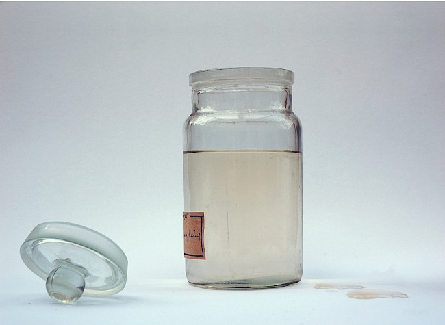
The closing off of anatomical collections happened in the nineteenth century, the age most other collections were opened up to the public. I wanted to figure out why. Hence, I studied how different audiences, both medical and nonmedical, used the collections – and on that morning in March, I stumbled on a quote that made me, literally, clap my hands in joy. It was in curator Richard Owen’s (1831, 7) report on his visit to the Parisian Muséum d’Anatomie Comparée: ‘Every preparation, used at lecture is taken out of the bottle to be demonstrated, and the same thing is done at the request of any scientific visitor who may wish to examine the part in a particular manner.’
This remark evoked such joy in me because it revealed how researchers and students worked with anatomical preparations: lids off and hands on. I had been suspecting this for a while. So far, however, most of my evidence was rather circumstantial. Owen’s explicit mention convinced me I was on the right track and stimulated me to systematically research the handling of preparations by both researchers and students. It turned out that active handling of specimens happened everywhere – not just in Paris, but also in London, Leiden, and other European cities. This transformed my idea of anatomical collections. I no longer understood them the way most medical historians do, as static objects, intended to be classified, arranged, and then looked at from a distance. Instead, I began to view them as dynamic, meant for active use: preparations were taken out of their jars, passed around in class to be felt and smelled, they were handled, reinvestigated, and even redissected. Anatomical collections, in short, were full of life. Grasping this lets us understand why, contrary to what is often assumed, anatomical collections remained relevant in the nineteenth century, when university medicine became increasingly hands on, with the birth of the clinic, the rise of the laboratory, and the advent of practical teaching.
My view of anatomical collections – and thus, my dissertation – had changed dramatically. And it would change even further, three months later, one balmy evening in May.
It was the first night of a three-day workshop on the afterlife of wet preparations, again at the Royal College of Surgeons. Medical historian Nick Hopwood delivered the keynote lecture, on embryological preparations. Hopwood reminded the audience that usually in the medical humanities we stress the similarities between models and preparations: both are artefacts – a fundamental assumption in my research project as well. But, he suggested, perhaps we should also investigate the differences between the two. As a starting point, he proposed an observation of the German philosopher of biology Hans-Jörg Rheinberger (2003, 2010, 232–43): although both anatomical models and anatomical preparations are made, only preparations are made of what they represent. Unlike a kidney model, a kidney preparation does not consist of wax or papier-mâché – it consists of what it represents: kidney.
Rheinberger’s observation became crucial for my argument, because it allowed me to explain not only why anatomical collections kept being used, but also why old, early modern collections kept being reused in new, nineteenth-century medicine. The preparations’ material properties enabled researchers to return to the original tissues again and again, extracting new information from them, a practice Rheinberger calls ‘epistemic recall’. Researchers could, for example, cut up macroscopic preparations to investigate their microscopic structure and thus adapt old preparations to new theories. Because of the practice, or at least the promise, of reuse, and because human remains were scarce, medical institutions held on to their preparations. Anatomical collections lingered in hospitals and laboratories, which moved to the outskirts of town, kept office hours, and, with professionalization, adopted a more closed atmosphere. Whereas other collections ended up in public museums, historicized, anatomical collections disappeared from the public view.
I had answered my original research question: anatomical collections disappeared from the public view because they, due to their material properties, never left the medical institutions that built them, and these institutions became increasingly inaccessible. To reach this answer, I had to learn to understand anatomical collections not as static artefacts, but as dynamic entities, made of what they represented. And, writing it all up, I slowly realized that this understanding was my most fundamental finding – it is also what makes me want to share my results with the readers of MAT.
For I hold that this understanding applies not just to nineteenth-century Leiden, but to other times and places as well, including ours. Collections of bodily material still flourish: tissue banks, frozen embryos, commercial cell lines. We tend to understand their contents as (commodified) artefacts, and assume it is technology that renders them immortal. Indeed, biotechnology prevents decay, but immortality requires more: ongoing relevance. The objects in human tissue collections have such relevance exactly because they are not fully artefactual – for although they are made, they are made of what they represent, be it cells, embryos, or diseased organs. This specific material property creates the eternal promise of reuse. It ensures bodily material is kept, not just for now, or for the next decades, but, as the Leiden anatomical collections show us, for the coming centuries – and perhaps, one might conclude, forever.
About the author
Hieke Huistra is a postdoctoral researcher at Utrecht University. She is interested in how medical practices and objects shape the way we experience our bodies. Her doctoral dissertation (‘Preparations on the Move: The Leiden Anatomical Collections in the Nineteenth Century’,Leiden University, 2013) shows how nineteenth-century anatomical collections became inaccessible to nonmedical audiences due to their material properties. She is currently revising this dissertation into a book titled Handling Anatomical Collections in the Nineteenth Century: Leiden and Beyond (to be published by Ashgate in 2016). In a new research project, she uses both digital and traditional historical methods to investigate ideas on fatness and overweight in early twentieth-century Dutch society.
References
Owen, Richard. 1831. Report to the Board of Curators of the Museum of the Royal College of Surgeons on the Museum d’Anatomie Comparée in the Garden of Plants, Paris. Owen papers, file MS0025/1/4/1. Royal College of Surgeons of England Library, London.
Rheinberger, Hans-Jörg. 2003. ‘Präparate–“Bilder” ihrer selbst: Ein bildtheoretische Glosse’. In Oberflächen der Theorie, vol. 1.2 of Bildwelten des Wissens: Kunsthistorisches Jahrbuch für Bildkritiek, 9–19.Berlin: Akademie Verlag.
Rheinberger, Hans-Jörg. 2010. An Epistemology of the Concrete: Twentieth-Century Histories of Life. Translated by G. M. Goshgarian. Durham, NC: Duke University Press.